Von Kossa Stain
The Von Kossa stain is a classical histological staining method primarily used to detect the presence of calcium salts in tissue sections, notably in pathological conditions like calcification.
Principle
The principle behind the Von Kossa staining technique relies on the ability of calcium salts to form an insoluble precipitate when exposed to silver nitrate under light. The silver is reduced to metallic silver by light, depositing a black or dark brown color wherever calcium salts are present in the tissue.
Method of Application
- Fixation: Tissue sections are fixed, commonly with formalin, to preserve structural integrity.
- Deparaffinization and Rehydration: Sections embedded in paraffin are cleared of paraffin and rehydrated through graded alcohol to water.
- Staining:
- The rehydrated sections are treated with a silver nitrate and exposed to bright light (usually UV) for 20-60 minutes. The light exposure is crucial as it catalyzes the reduction of silver nitrate to metallic silver in areas where calcium salts are present.
- After exposure, the slides are washed in distilled water to remove unreacted silver.
- Counterstaining (Optional):
- To enhance contrast and detail, sections may be counterstained with nuclear fast red or another mild counterstain, which colors the non-mineralized tissue components, making the black mineral deposits stand out more prominently.
- Washing and Mounting:
- The stained sections are then rinsed, dehydrated through a series of alcohols, cleared in xylene, and mounted under a coverslip with a permanent mounting medium.
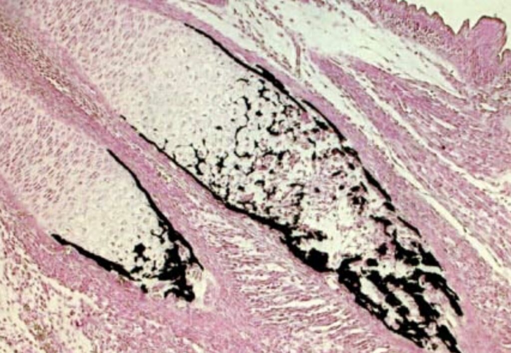
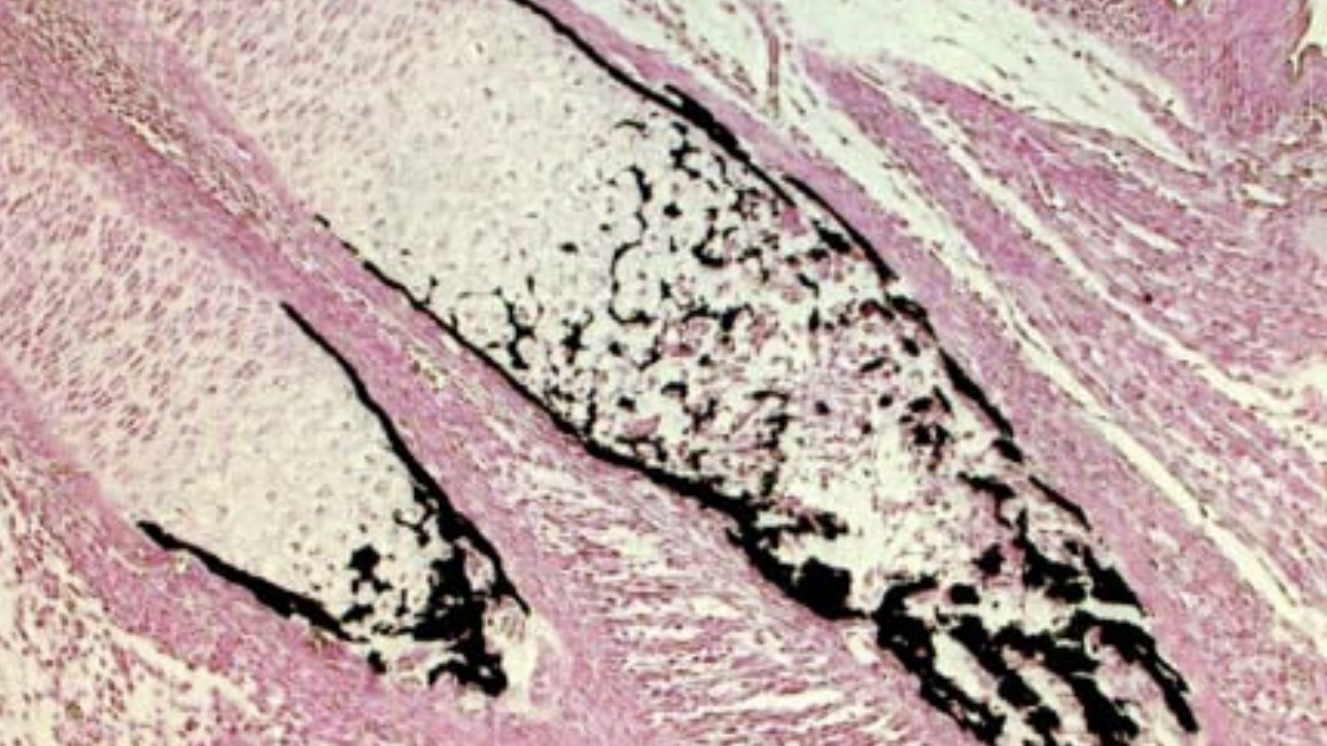
This microscopic image demonstrates the Von Kossa staining method:
- Black Staining: Indicates the presence of calcium deposits within the tissue, stained black by the reduction of silver under light exposure.
- Pink Background: Non-mineralized tissue components are counterstained with nuclear fast red, providing a clear contrast highlighting calcium deposits’ distribution and morphology.


Leave a Reply