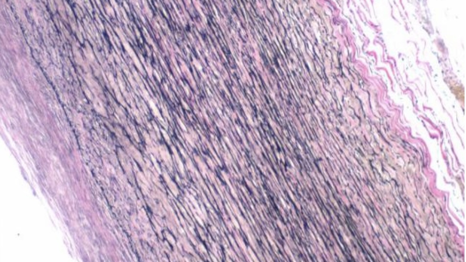
Verhoeff-van Gieson Stain
The Verhoeff-van Gieson (VVG) stain is a specialized histological staining method used primarily to examine tissue elastic fibers. Here’s a detailed look at how it works and its application:
Principle
The VVG stain utilizes the properties of iron hematoxylin and van Gieson’s picro-fuchsin to selectively highlight elastic fibers, collagen, and other connective tissue elements:
- Iron Hematoxylin: This component stains elastic fibers, cell nuclei, and other tissue elements black.
- Van Gieson’s Picro-Fuchsin: This is a mixture of picric acid and acid fuchsin that stains collagen fibers red and other connective tissues a yellow or pale pink.
Method
The VVG staining procedure involves several key steps:
- Fixation: Tissues are fixed, typically in formalin, to preserve their morphology.
- Deparaffinization and Rehydration: If the tissue is embedded in paraffin, the sections are cleared of paraffin and rehydrated through a graded alcohol series to water.
- Staining with Iron Hematoxylin:
- The sections are stained with iron hematoxylin, which imparts a black color to elastic fibers and nuclei.
- The slides are then washed and differentiated in a ferric chloride solution to enhance the specificity of the staining for elastic fibers.
- Counterstaining:
- After rinsing, the sections are stained with van Gieson’s picro-fuchsin solution. This stains collagen fibers red and provides a visual contrast to the black-stained elastic fibers.
- Dehydration and Mounting:
- The stained sections are dehydrated through alcohols, cleared in xylene, and mounted with a resinous medium.
Utilization
VVG staining is extensively used in pathology to:
- Evaluate the integrity and distribution of elastic fibers in tissues, crucial in diagnosing diseases affecting the vascular system and connective tissue disorders.
- Differentiate between connective tissue elements in complex tissues, helping assess pathological changes involving the elastic and collagenous components.
This method is valuable in clinical diagnostics and research settings, providing crucial insights into tissue structure and pathology.
Visualization
Under a microscope, elastic fibers stained by the Verhoeff-van Gieson method appear black, collagen appears red, and other tissues have a yellow or pink hue. This contrast is vital for detailed tissue analysis, particularly in diagnosing connective tissue and vascular system diseases.
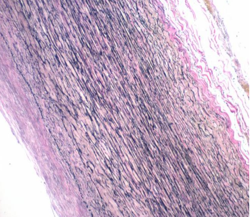
- Elastic fibers: Stained blue-black to black, highlighting their detailed network within the aortic wall.
- Nuclei: Appear blue to black, indicating cellular structures.
- Collagen: Stained red, contrasting with the elastic fibers, illustrating the supportive tissue structure.
- Other tissue components: Stained yellow, displaying the background matrix and non-elastic, non-collagenous elements.


Leave a Reply