The Gram Stain
The Gram stain is a fundamental technique used in microbiology to differentiate bacteria into two large groups based on their cell walls’ chemical and physical properties.
Principle
The principle behind Gram staining is based on the ability of bacterial cell walls to retain the crystal violet dye during solvent treatment. Bacteria can be classified as:
- Gram-positive: These bacteria have a thick peptidoglycan layer that retains the crystal violet dye, appearing blue or purple.
- Gram-negative: These bacteria have a thinner peptidoglycan layer and an outer membrane that does not retain the crystal violet dye when washed with a decolorizer such as ethanol or acetone. They are counterstained with safranin or fuchsin, appearing red or pink.
Method of Application
The standard Gram staining procedure involves several steps:
- Fixation: Bacterial smear is heat-fixed on a slide to kill and adhere the bacteria to the slide.
- Staining with Crystal Violet:
- The slide is flooded with crystal violet dye and left for about one minute.
- After staining, the slide is gently rinsed with water.
- Mordant Application:
- Iodine solution, which acts as a mordant, is applied to the slide. This step helps to form a crystal violet-iodine complex that is more difficult to wash out of the cell walls.
- The slide is rinsed again after a minute.
- Decolorization:
- A decolorizing agent (either alcohol or acetone-alcohol) is briefly applied to the slide. This step is critical as it removes the dye from Gram-negative cells but not from Gram-positive cells.
- The slide is quickly rinsed with water to stop the decolorization process.
- Counterstaining:
- A contrasting dye, typically safranin, is applied to the slide, staining the Gram-negative bacteria for 30 seconds to one minute.
- The slide is then rinsed and dried.
- Microscopic Examination:
- The slide is examined under a microscope to determine the color of the bacteria, thus classifying them as Gram-positive or Gram-negative.
Utilization
Gram staining is widely used in:
- Clinical diagnostics to quickly identify and categorize bacterial infections.
- Microbiology research to study bacterial taxonomy and morphology.
- Antibiotic susceptibility testing where it provides initial insights into the type of antibiotic that may be effective against a particular bacterium.
This method is crucial for rapidly identifying and characterizing bacteria in both medical and research settings.

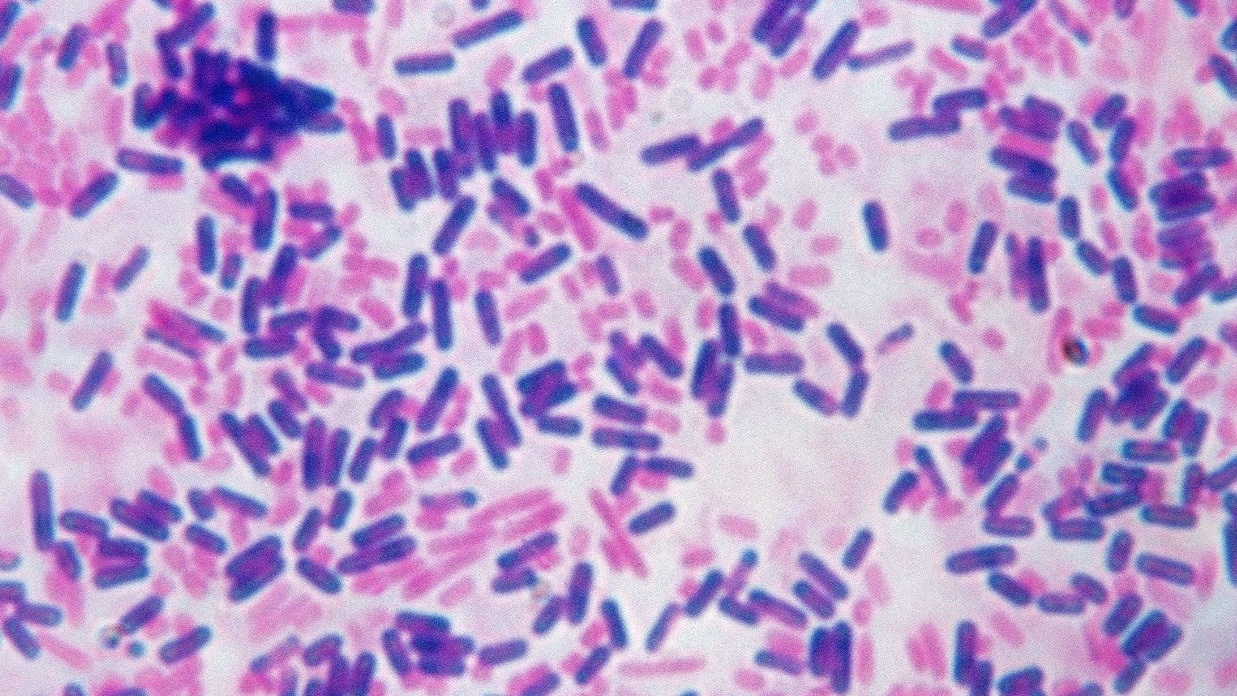
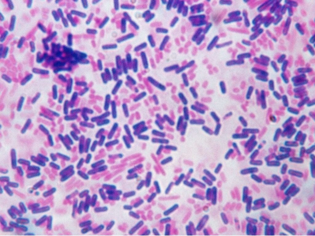
This microscopic image displays the differential staining characteristic of the Gram staining technique:
- Purple Cells: Represent Gram-positive bacteria, characterized by a thick peptidoglycan layer that retains the crystal violet dye, appearing purple.
- Pink Cells: Indicate Gram-negative bacteria, which have a thinner peptidoglycan wall and do not retain the primary stain but are counterstained pink by safranin.


Leave a Reply