Special Stains
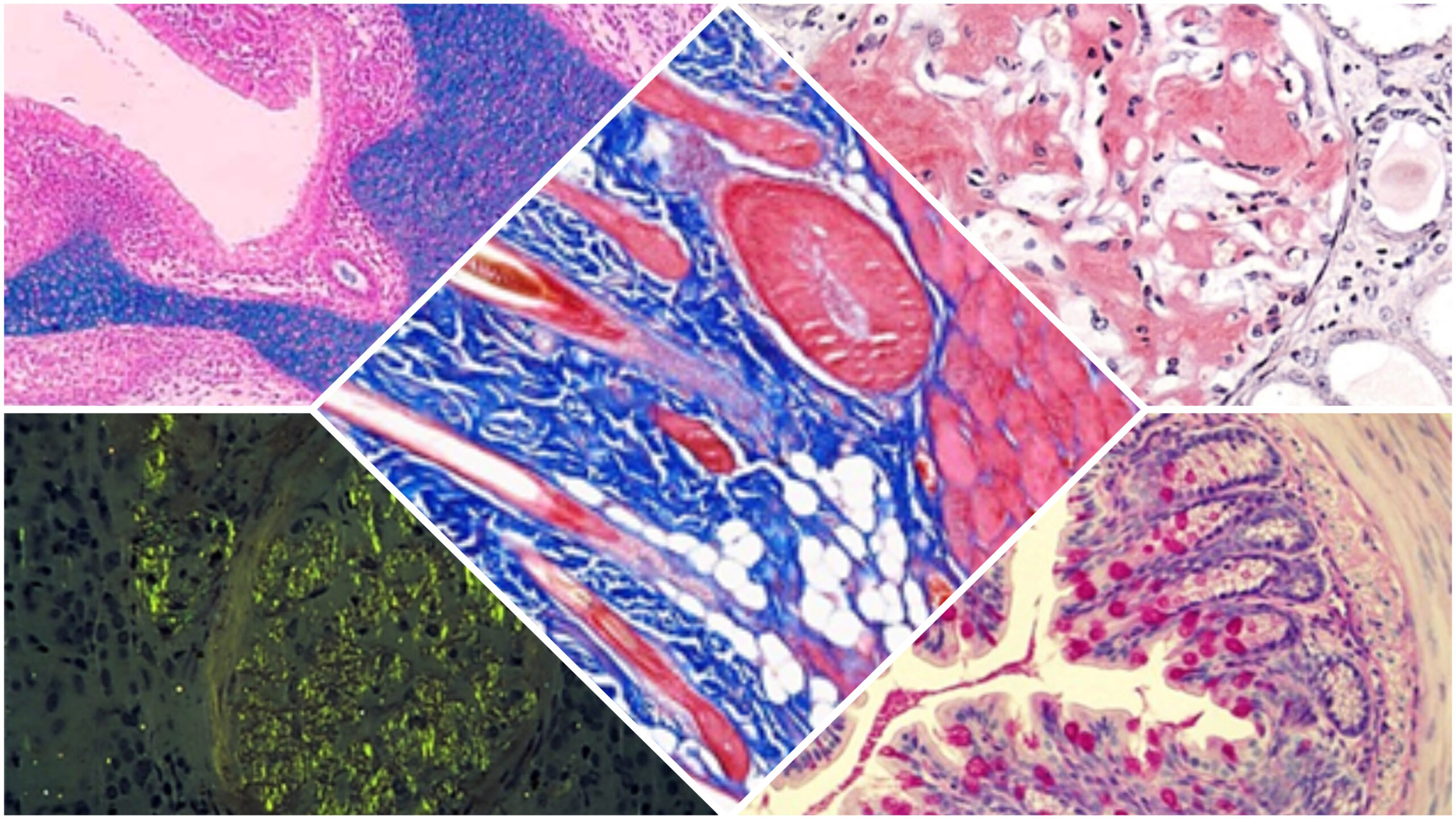
Special stains are used to highlight specific tissue or cellular components in histopathology. The hematoxylin and eosin (H&E) stain is the primary screening test. Typical details appear clearly on H&E, while abnormalities based on morphology may require further testing with special stains. These stains are classified by their targets, including carbohydrates, amyloids, connective tissue, microorganisms, minerals, and pigments.
Histology Special Stains Classification
Special histology stains emphasize particular tissue or cellular components that are challenging to discern using the standard hematoxylin and eosin (H&E) stain. This classification categorizes the stains according to their targeted staining preferences. The most crucial classifications are presented here:
1. Carbohydrates:
- Periodic Acid-Schiff (PAS): Stains glycogen, mucins, and basement membranes.
- Alcian Blue: Stains acidic mucins and glycosaminoglycans.
2. Amyloids:
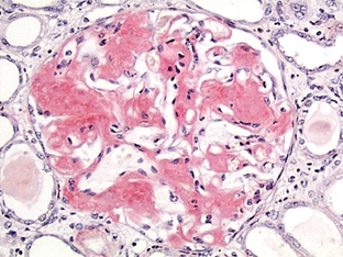
- Congo Red: Stains amyloid deposits showing apple-green birefringence under polarized light.
3. Connective Tissue:
- Masson’s Trichrome: Differentiates between muscle, collagen, and fibrin.
- Verhoeff-van Gieson (VVG): Stains elastic fibers.
4. Microorganisms:
- Gram Stain: Differentiates between Gram-positive and Gram-negative bacteria.
- Ziehl-Neelsen (Acid-Fast) Stain: Identifies acid-fast bacteria like Mycobacterium.
- Grocott’s Methenamine Silver (GMS): Stains fungi and certain bacteria.
5. Minerals and Pigments:
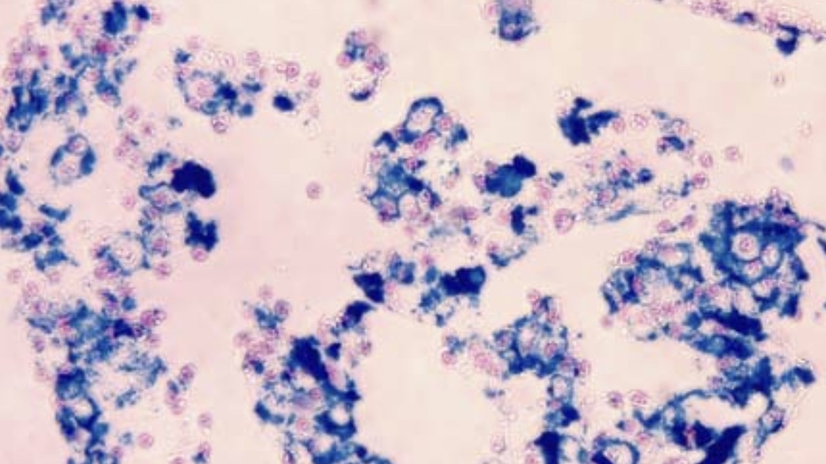
- Perl’s Prussian Blue: This stain uses iron salts to detect iron deposits in tissues, staining them blue.
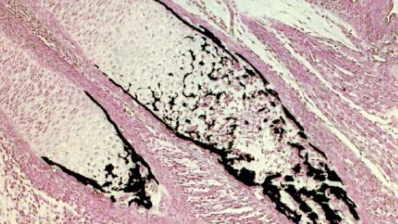
- Von Kossa: Stains calcium deposits.
These special stains provide additional diagnostic information by targeting specific components within the tissue, aiding in identifying and characterizing various pathological conditions.


Leave a Reply