Skip to the content
al-rafiah.comlogo darklogo light
  • Home
  • About Us
  • Education
  • Services
    • Digital Pathology
    • Histology Services
    • Academic Writing
    • Statistical analysis
    • Consultation
    • Workshops and Lectures
  • Blog
  • Publication
  • My Book
  • Contact Us
Search
al-rafiah.com
  • Home
  • About Us
  • Education
  • Services
    • Digital Pathology
    • Histology Services
    • Academic Writing
    • Statistical analysis
    • Consultation
    • Workshops and Lectures
  • Blog
  • Publication
  • My Book
  • Contact Us

Special Stain

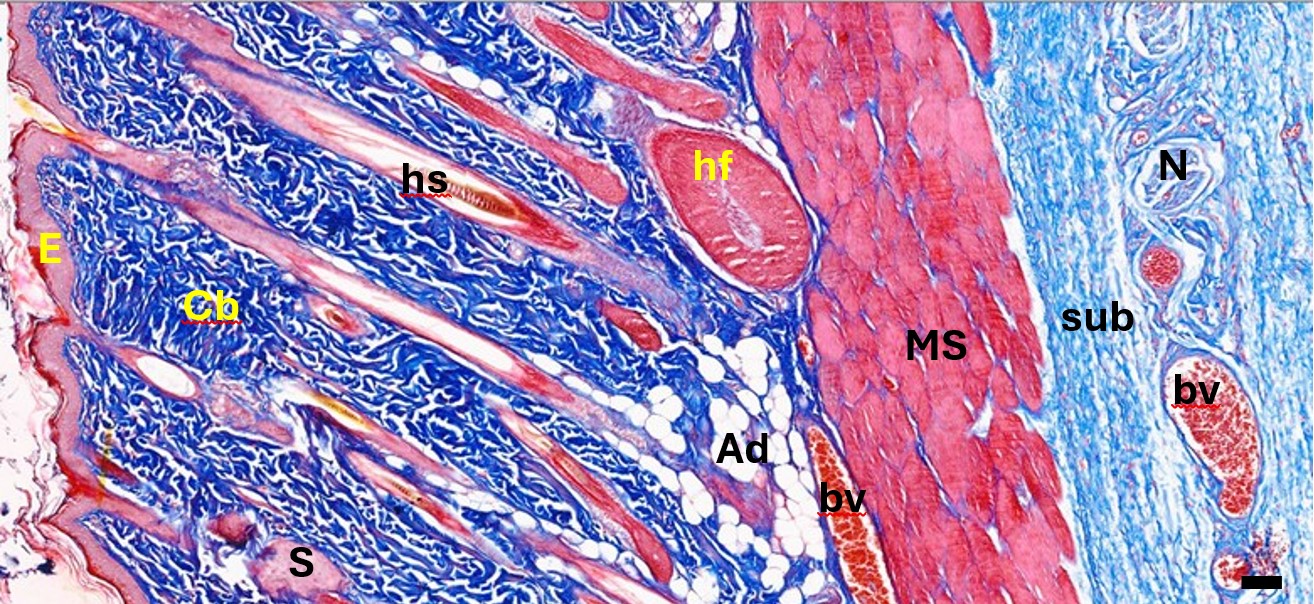
Figure 1: Photomicrograph of normal back skin of one of the experimental rats showing the cutaneous layers and their contents. Epidermis (E), collagenous bundles (Cb), hair shaft (hs), Hair follicle (hf), sebaceous gland (S), adipose tissue (Ad), blood vessels (bv), nerves (N), cutaneous muscle (MS) and subcutaneous tissue (sub). Magnification: X40. Stain: MS. Bar: 50 µm.

Masson’s trichrome :

Date:

November 10, 2022

Category:

Histopathology
Previous Project View All Next Project
Histomorphometry Studies

Al-rafiah

  • About Us
  • Team
  • How we Work
  • Careers

Services

  • Digital Pathology
  • Histology Service
  • Academic Writing
  • Statistical Analysis
  • Consultation
  • Workshops and Lectures

Contact

  • Send us a Message
  •   +966 50 593 1745
  •   dralrafiah@gmail.com

© 2024 Al-rafiah., All Rights Reserved

Back to top Drag