Periodic Acid-Schiff (PAS)
The PAS Diastase stain diagnoses polysaccharide-related disorders, such as glycogen storage diseases. Diastase (α-amylase) is employed to depolymerize glycogen into smaller sugar units in the form of glucose or maltose. These monosaccharides are then washed away from the tissue sections. Typically, two slides are prepared for each case: one undergoes diastase digestion, while the other does not. Both slides are then stained with PAS. The slide treated with diastase will not show PAS staining due to the breakdown and removal of polysaccharides.
Step 1: Preparation
- Deparaffinization: Tissue sections are deparaffinized by immersing them in xylene and rehydrated through a series of graded alcohols in water.
- Oxidation: The tissue sections are treated with periodic acid, which oxidizes the polysaccharides, creating aldehyde groups.
Step 2: Staining
- Schiff Reagent Application: The sections are then treated with Schiff reagent, which reacts with the aldehyde groups to form a magenta or pink color, indicating the presence of polysaccharides.
- Washing: The slides are washed in running tap water to remove excess Schiff reagent and develop the magenta color.
Step 3: Counterstaining (Optional)
- Counterstaining: Hematoxylin may be used as a counterstain to provide contrast by staining the nuclei blue.
- Dehydration and Clearing: The tissue sections are dehydrated through graded alcohols, cleared in xylene, and mounted with a coverslip.
PAS-Diastase Method (for Glycogen)
In cases where glycogen detection is specifically needed, the PAS-Diastase method is used:
- Diastase Treatment: One of the tissue sections is treated with diastase (α-amylase) to digest glycogen into smaller sugar units, which are then washed away.
- PAS Staining: The digested (diastase-treated) and undigested slides are stained using the PAS method.
- Comparison: The digested slide will lack PAS staining in areas where glycogen was present, as it has been removed by diastase treatment. The undigested slide will show positive PAS staining in areas with glycogen.
The PAS method is a valuable tool in histology for visualizing and diagnosing conditions related to polysaccharides and mucosubstances in tissues.
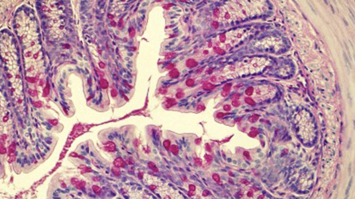
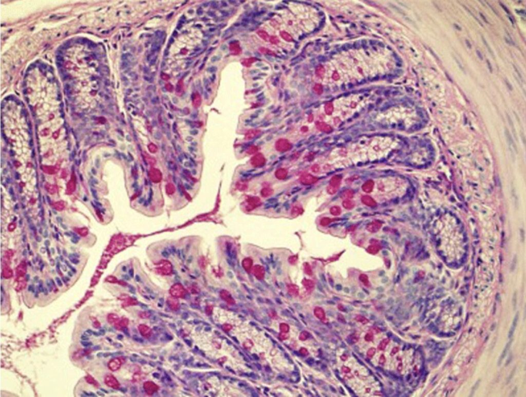
The Periodic Acid-Schiff’s (PAS) stain detects polysaccharides, mucosubstances, and glycogen in tissues. It produces distinct colors that highlight these components:
- Magenta/Pink: Polysaccharides, glycogen, mucosubstances, and basement membranes stain magenta or pink due to the reaction between Schiff reagent and aldehyde groups formed by periodic acid oxidation.
- Blue/Purple (optional): Nuclei may be counterstained with hematoxylin, which stains them blue or purple, contrasting the magenta/pink-stained structures.
These colors help identify and diagnose various tissue components and pathological conditions.


Leave a Reply