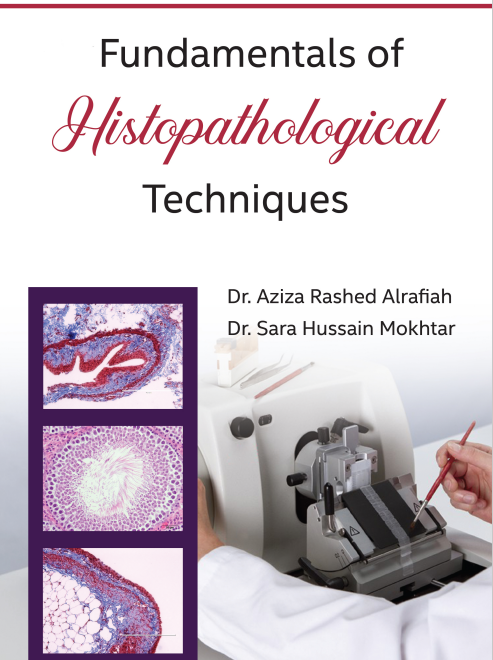
Fundamentals of
Histopathological Techniques
The Fundamentals of Histopathological Techniques is a comprehensive guide to preparing histological slides, covering procedures, staining protocols, and safety measures. Ideal for students, technologists, researchers, and pathologists, it ensures precise and efficient laboratory practices.

Preface to the first edition
Histological techniques have been expanded to include a wide range of disciplines. As a result, the level and breadth of knowledge required by trainee examiners in histology and histopathology technology have increased dramatically.
A detailed book on histological techniques that is appropriately reliable in various of domains that a technologist require is needed. Immunohistochemistry and immunofluorescence play crucial roles in inpatient treatment and have well-defined diagnostic and screening functions with quality assurance standards. In situ hybridization is considered an advanced technique to localize specific DNA in each specimen on tissue or cells. Many books focus on one topic, and the specialized technician should read them as part of their self-education. Therefore, a book covering the complete range of histotechnology, from the basics of tissue fixation and the formation of paraffin sections to the more complex concepts of electron microscopy, is now necessitated.
Thus, our goal was to create a book that is considered a detailed reference for those who are preparing for histopathology exams nationally and internationally. In addition, the book will be helpful for more advanced students, researchers, histologists, and pathologists. It covers all the fundamental laboratory procedures used in histology and histopathology labs, including their principles and methods. The contributors have taken care to provide, if appropriate, the theoretical background of the techniques. Several graphs and illustrations have been used to explain how the processes work. Along with various advanced techniques, discussions have also included microscopy and lab quality control.
We hope we have created a modern book that will be useful to all histopathology graduate students, practicing histopathologists, and lab technicians worldwide.
Prof. Aziza Rashed Alrafiah
King Abdulaziz University, Saudi Arabia
Chapter 1
Introduction
Detailed Table of Contents
This chapter presents a brief account of the origin of histology and histopathology. In fact, after the invention of microscopes, the study of delicate tissue details became possible; in addition, studying histochemistry allowed the use of different stains and the characterization of different tissues and cells. Both histologists and pathologists have utilized the unlimited progress in histotechnology. With histopathological techniques, physicians have a reliable source of disease diagnosis. Nevertheless, optimal technical procedures and safety measures must be undertaken to obtain correct results safely.
Chapter 2
Laboratory Quality Management-Overview
In this chapter, the Sound Quality management system (QMS) ensures that laboratories meet all needs. Patient care is an integral part of QMS, and meeting patients’ needs is a primary goal of the laboratory. The principal element for a successful QMS is Management commitment. Quality System Essentials (QSEs) must be well-defined and documented. The quality control process is essential to identify and remove mistakes before reporting patient results.
Chapter 3
Laboratory Safety
Histopathology lab work is fascinating, but it must be attended to carefully by observing the dangers inside, microtome knives, glassware, chemicals, biological substances, etc. To avoid personal injury and property damage, all workers and employees in the lab must be aware of their surrounding environment and adopt safety measures.
Chapter 4
Light Microscopy
Chapter 4 discusses the light microscope, considered one of the essential equipment in histological and histopathological investigations and lab diagnosis. Therefore, it is worthwhile for all lab workers and researchers to know enough about the theory and practice of light microscopy and the proper use of the light microscope. This chapter added valuable details about the light microscope and its use.
Chapter 5
Source of Tissue Samples
In this chapter, tissue sampling is essential to reaching good results and the correct diagnosis. The tissue specimen must be fresh, of considerable size, and 0.5 cm or less in one of its dimensions; it must be immediately immersed in a suitable fixative solution. Samples must take an accession number and be sent directly to the histopathology lab for processing.
Chapter 6
Tissue fixation
In this chapter, tissue fixation is explained. Fixation aims to preserve tissue and cell components in a state that simulates actual living conditions by stopping post-mortem autolysis and preventing microbial contamination. The immersion method is commonly used in which specimens are immediately immersed (1 volume of specimen: 10 volumes of fixative). Fixatives are divided into five primary categories: aldehydes, alcohols, picrates, mercury, and oxidizing agents.
Chapter 7
Grossing
Chapter 7 contains concise notes about macroscopic examination and trimming of the specimen to render it ready for processing and sectioning (grossing). Sections can be obtained directly from it if valuable lessons are exposed near the paraffin block face. In addition, cassettes must be well-defined and contain considerable parts of the specimen.
Chapter 8
Tissue Processing
This chapter presents the tissue processing schedule in a simplified way. As the fixed specimen is not yet ready for sectioning, it must be impregnated in an impregnation medium to make it easy to be thin-sectioned. Tissue processing begins with dehydration (the most used dehydrate the ethyl alcohol) followed by clearance, a process achieved by an organic solvent such as xylene. Then, the specimens are impregnated in paraffin wax which is easy to be sectioned by microtome knives.
Chapter 9
Embedding
In this chapter, a brief account of the embedding process is added. Embedding in histopathology labs aims to form tissue blocks easily sectioned by microtomes. The most used embedding technique is the paraffin wax technique; however, the cryostat's freezing technique during surgical operations and sectioning is sometimes used to obtain rapid results and decisions. During the formation of the tissue block, specimens must be oriented correctly in the mold to gain sections from the lesions or the chosen tissue areas.
Chapter 10
Microtomy
This chapter presents an account of microtomy. Translucent tissue sections must be obtained by microtome and mounted on a glass slide to be examined by the microscope. There are several microtomes, of which the rotary microtome is the most used. In this chapter, the structure and manual use of the rotary microtome is presented; in addition, common sectioning artifacts and troubleshooting are also listed.
Chapter 11
Theory of Staining
In this chapter, the theory and practice of tissue staining are stated. Different types of stains are recorded. Staining protocol using hematoxylin to stain the nuclei and eosin to stain cytoplasm is the commonly used stain in histopathological labs; however, different specific stains are also present. Mounting the stained section in a translucent medium protects and saves it for a long time.
Chapter 12
Special Stains
In this chapter, detailed notes about specific stains are conducted. Special stains can be classified according to the staining target, including carbohydrates, amyloids, connective tissue, microorganisms, minerals, and pigments. The chemical reactivity of tissue and cellular components allow the staining and localization of many of them. As a result, many protocols of special stains are now applicable in histopathological labs for diagnostic purposes.
Chapter 13
Immunohistochemical techniques
In this chapter, the antigen-antibody reaction is used in an advanced branch called immunohistochemistry, in which specific antigens could be localized in each tissue by a labelled antibody. The label may be enzyme as peroxidase or fluorochrome as fluorescein; light and fluorescent microscopes are used to examine tissue antigens. The theory of immunohistochemistry and immunohistochemical techniques are also presented in this chapter.
Chapter 14
Fluorescent In Situ Hybridization (FISH)
This chapter explains in situ hybridization as an advanced technique to localize a specific DNA in a given specimen, either on tissue or cells. First, the fluorescent in situ hybridization (FISH) method is briefly presented. However, FISH is a sensitive technique and time-consuming, so it is used as a research technique rather than for laboratory diagnosis.

