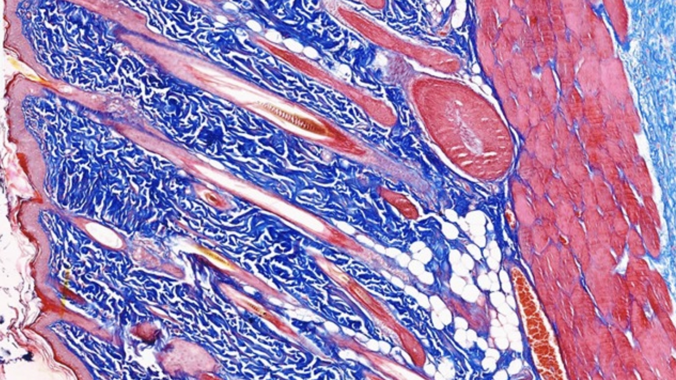
Masson’s Trichrome Stain
The Masson’s Trichrome stain is a common histological staining technique used to differentiate between collagen and muscle fibers in tissue samples.
Principle
Masson’s Trichrome stain utilizes a combination of dyes to stain various components of the tissue differentially:
- Aniline Blue: Stains collagen fibers blue.
- Biebrich Scarlet-Acid Fuchsin: Stains muscle fibers, cytoplasm, and keratin red.
- Weigert’s Hematoxylin: Typically used to stain nuclei black.
This staining technique is beneficial for identifying increases in collagenous tissue (fibrosis) and for detailed examination of muscle pathology.
Method of Application
The Masson’s Trichrome staining process involves the following steps:
- Fixation: Tissue samples are fixed, typically with formalin, to preserve the tissue architecture.
- Deparaffinization and Rehydration: Paraffin-embedded tissue sections are cleared of paraffin and rehydrated through a series of graded alcohols to water.
- Staining:
- Nuclear Staining: Sections are stained with Weigert’s hematoxylin to stain nuclei black.
- Rinsing and Staining for Muscle: After rinsing, sections are stained with Biebrich Scarlet-Acid Fuchsin to stain muscle fibers red.
- Differentiation: Sections are differentiated in phosphomolybdic-phosphotungstic acid to remove excess red stain except in muscle fibers.
- Collagen Staining: Finally, sections are stained with aniline blue to stain collagen blue.
- Dehydration and Clearing:
- Sections are dehydrated in alcohols, cleared in xylene, and mounted under a glass coverslip with a resinous medium.
Utilization
Masson’s Trichrome stain is widely used in pathology for:
- Muscle Tissue Analysis: To assess muscle damage or disease.
- Fibrosis Evaluation: To detect and quantify fibrotic changes in tissues such as the liver, kidney, and lungs.
- Tumor Differentiation: Helps differentiate between tumor tissue and surrounding stroma.
This method is precious in clinical diagnostics and research because it can reveal detailed tissue structural differences.
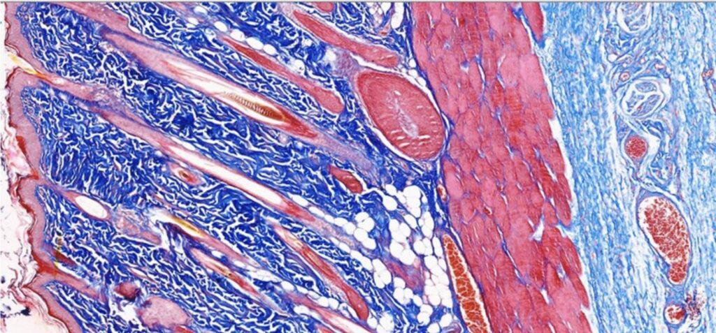
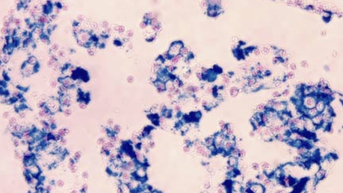
This image illustrates the differential staining of skin components using Masson’s Trichrome technique:
- Nuclei: Appear dark blue to brown, clearly delineated to highlight cellular structures.
- Cytoplasm: Stained red, providing a stark contrast that aids in identifying cellular bodies within the tissue.
- Collagen: It appears blue surrounds the cellular structures and fills the extracellular matrix, indicative of the collagenous connective tissue framework within the skin.


Leave a Reply