Grocott Methenamine Silver Stain
The Grocott’s Methenamine Silver stain, or Gomori methenamine silver stain, is a histological staining technique used primarily to detect fungal organisms and other pathogens in tissue sections.
Principle
The Grocott Methenamine Silver (GMS) stain relies on the ability of fungal cell walls and other organisms to bind silver. The staining process involves reducing silver methenamine, where the silver is deposited selectively in fungal walls and some other structures, making them appear black under a light microscope. This contrast against the lightly stained background tissues allows for a clear visualization of the presence and morphology of the pathogens.
Method of Application
The GMS staining procedure typically follows these steps:
- 1. Fixation: Tissue samples are fixed, usually with formalin, to preserve the structural integrity of the tissue.
- 2. Deparaffinization and Rehydration: Sections embedded in paraffin are deparaffinized in xylene and rehydrated through graded alcohols to water.
- 3. Silver Staining:
- Sections are treated with chromic acid for oxidation, which enhances silver staining.
- They are then washed and treated with a methenamine silver solution. During this step, silver ions bind to the fungal structures.
- The sections are heated, accelerating the silver deposition on the fungal cell walls.
- 4. Toning:
- After rinsing, the sections are toned with a gold chloride solution, which reduces the silver to a visible metallic silver state.
- 5. Counterstaining:
- Sections are counterstained with light green or another mild counterstain to provide a contrast, making the black-stained fungi more prominent against a green or pale background.
- 6. Dehydration, Clearing, and Mounting:
- The slides are dehydrated in graded alcohols, cleared in xylene, and mounted under a glass coverslip.
Utilization
GMS staining is used extensively in medical pathology to:
- Detect Fungi: Particularly useful in diagnosing fungal infections, such as Pneumocystis pneumonia, aspergillosis, and other systemic or localized fungal diseases.
- Identify Pneumocystis jirovecii, Especially in lung tissue samples from immunocompromised patients.
- Detect Some Bacteria and Protozoa, Including Legionella and Histoplasma.
This method is crucial for accurately diagnosing infections, particularly in clinical settings where rapid and accurate pathogen identification is vital.

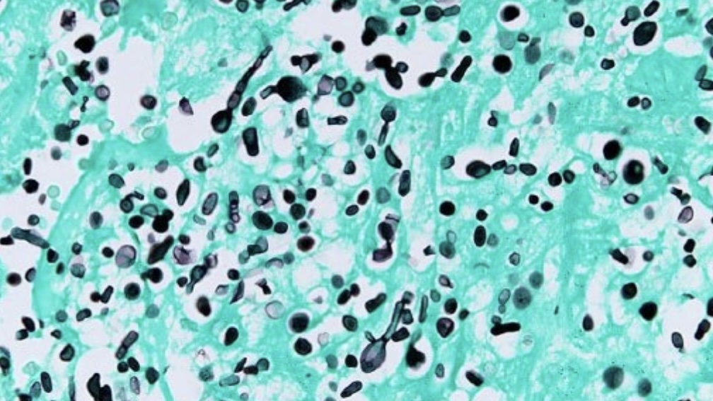
This microscopic image showcases the specialized application of the Grocott Methenamine Silver staining technique:
- Black Structures: These highlight fungal elements within the tissue, which absorb the silver stain due to the unique properties of their cell walls and appear distinctly black.
- Background Tissue: Lightly stained, providing a clear contrast that emphasizes the presence and morphology of the fungal structures.


Leave a Reply